190斤胖胖糖尿病患者危重,因背部疖子长到2000 cm²恶臭进ICU,伤口愈合仍遥遥无期
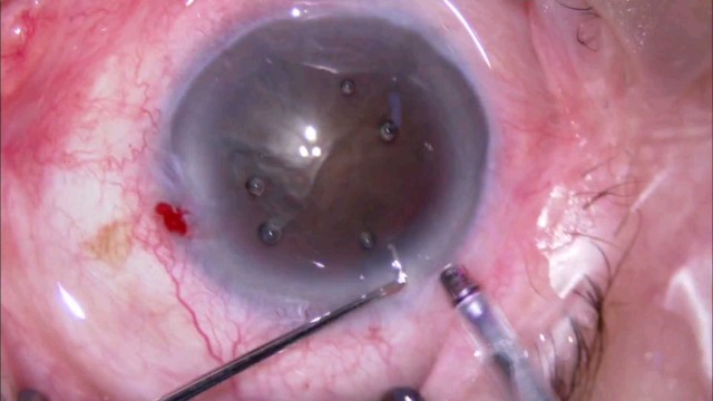
初看照片,就是背部一个巨大痈,基底肉芽红润,应该不难治疗:患者躺下,我换药时,进一步评估,才发现这个背部痈是到目前为止我接诊面积最大的、感染潜行最深,预计治疗时间也是最长的:创面大小:30*15cm,四周潜行约50*40cm进一步探查:四周还有坏死和流脓进一步沟通得知:一个月前就诊上级医院时,血糖爆表,血糖仪测不出,抽血38.8mmol/L,急诊手术后就住进ICU;五天后病情稳定,再次清创手术,才转出ICU,后续就是反复多次清创换药,大家可以想想早期治疗过程多么恶臭;早期的照片:(主管医生提供)患者能活命,真得好好感谢上级医院诊治的医生和相关科室,尤其是每天换药的那位医生,忍受一个半小时的臭味